A LABORATORY AIMED AT MAKING A DIFFERENCE. Through research, discovery, and innovation we shape the future of nanotechnology, biotechnology, health and medicine…
SYNTHETIC CAPILLARIES

LASER GUIDED ASSEMBLY OF METAMATERIALS
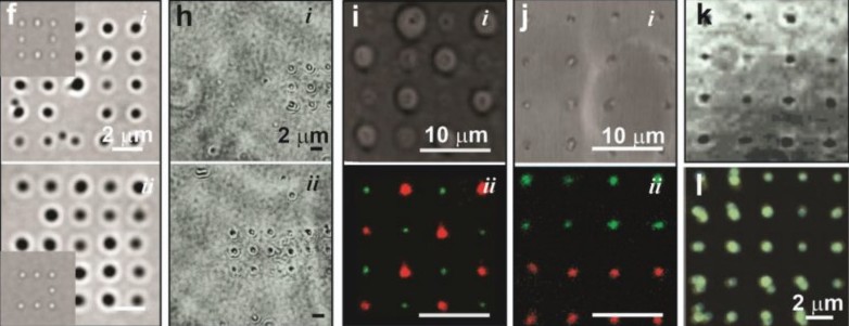
Above: Modular Assembly of 2D lattices of NPs using light gradients
PROTEIN SEQUENCING WITH A SOLID- STATE NANOPORE


Above: (Top, left) A TEM image taken in situ of a pore immediately after sputtering through an a-Si membrane; (center) MD simulation of A𝛽1-42 transiting a sub-nanopore; (right) Bi-conical topography of nanopore. (Bottom, left) A schematic cutaway of a single molecule biotin-A𝛽1−42 peptide, tethered to the tip of an AFM cantilever; (center) Concomitant measurements of the force and current are shown as a single, biotin-A𝛽1−42 molecule was pulled once, through a sub-nanopore;(right) A kymograph of the AC current is shown that represents a compilation of auto-correlation functions.
SINGLE CELL SECRETIONS USING A NANOPORE

Above (left) A schematic of the microfluidic cell conveyer and optical tweezers is shown; (b) A drawing of the seven-port microfluidic device with a nanopore embedded in the cross-bar is shown; (c) A false-color (perspective) reconstruction is shown of an MDA-MB-231 cell (green) suspended over a silicon nitride membrane with a nanopore in it. Inset: A transmission electron micrograph (TEM) of a (2.8 nm × 2.9 nm →) 6.4 nm2 cross-section nanopore is shown; the shot noise highlighted by the dashed circle delineates the pore.
LIVE BACTERIAL PHYSIOLOGY WITH STEM

STEM imaging of live bacterial physiology with a liquid cell
Above: (left) Image of negatively stained P1 phage on silicon nitride; (center) P1 phage adhering to K12 E. coli after staining; (right) The corresponding line scans that were used to evaluate the contrast and resolution .