
Where thoughts sparkle like fish in a sea of neurons.
Inside your brain, a coral reef of activity is always at work. Neurons light up as you think, move, and feel. These cells connect like glowing sea creatures in an electric current. Some provide support, others carry signals, but all work together to create your unique mind. These images, from fruit flies to human brains, show the beauty of how we think.
Try this: Can you spot the “spark” of a thought? What might this brain cell be thinking? If your thoughts swam like fish in a reef, what kind would they be?

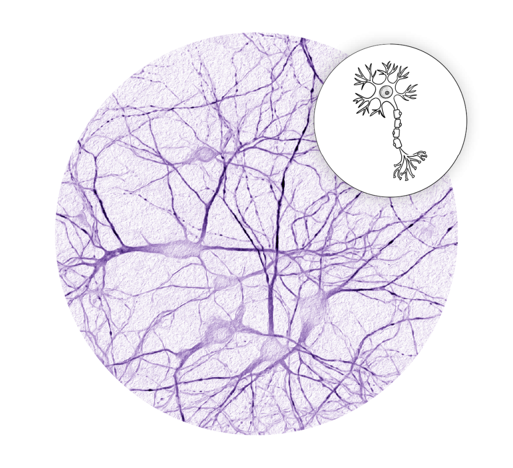
Neural Network: Human neurons (Credit: Dr. Louis Dang)
Bright green threads spread and branch like coral. These are brain cells reaching out to connect. Neurons pass signals to help us think, move, and remember, so these tiny links are the basic “wiring” that makes the brain work. Understanding how these networks behave, especially in cases of genetic epilepsy, can help researchers design more effective treatments for seizure disorders.
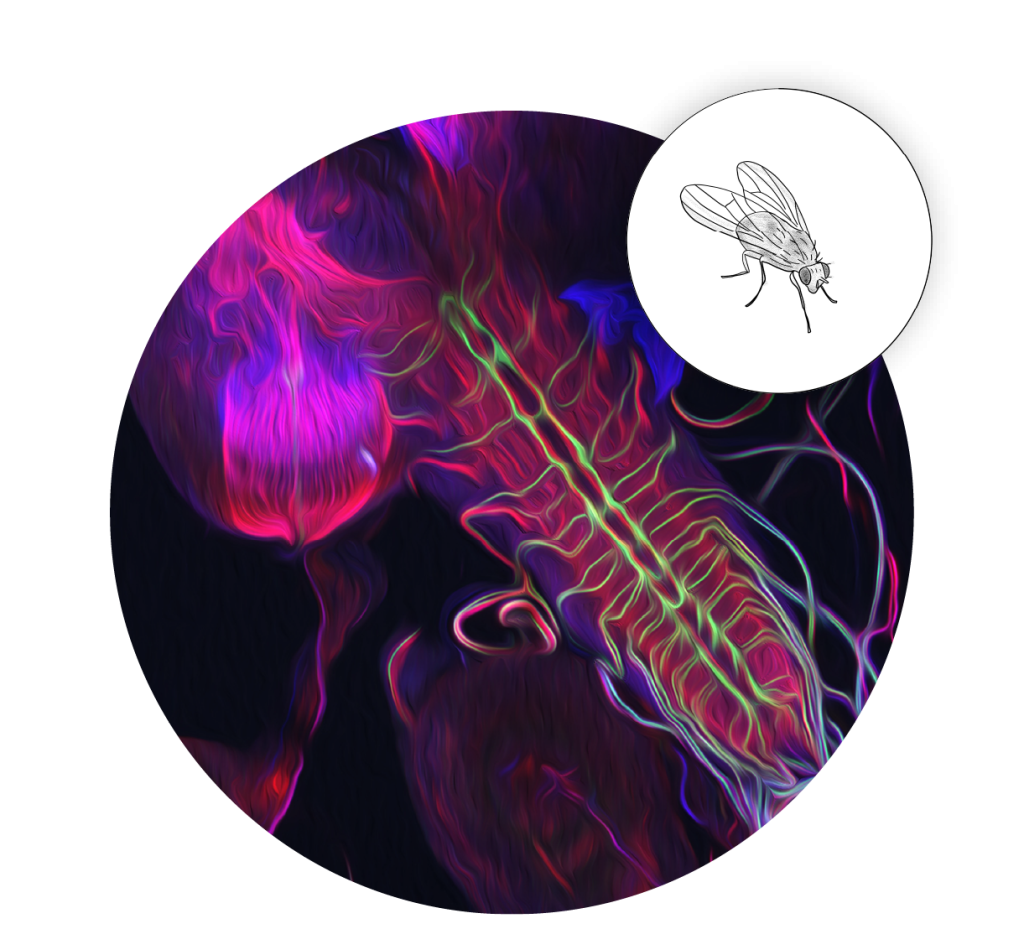
Fly Control: Nervous system of fruit fly larva (Credit: Dr. Bing Ye)
In this image of the nervous system of a fruit fly, nerves are highlighted in red, while green marks special neurons that respond to harmful stimuli, glowing like alarms. Researchers recently identified a protein in these green neurons, called DSCAM (Down’s Syndrome Cell Adhesion Molecule), that shapes how they connect with neighbors. Because DSCAM is also present in humans, studying it in fruit flies helps us better understand its role in human neurological conditions like Down syndrome.
Hippo Crosstalk: Neurons in a Petri Dish (Credit: Dr. Pilar Rivero-Ríos)
The hippocampus, named after the Greek word for seahorse, is a region of the brain critical for memory, emotional response, and spatial navigation. In this image, the branching projections of the neurons form connections (synapses) that allow them to communicate. Studying how these cells signal each other in the lab helps scientists better understand how the brain processes and stores information.
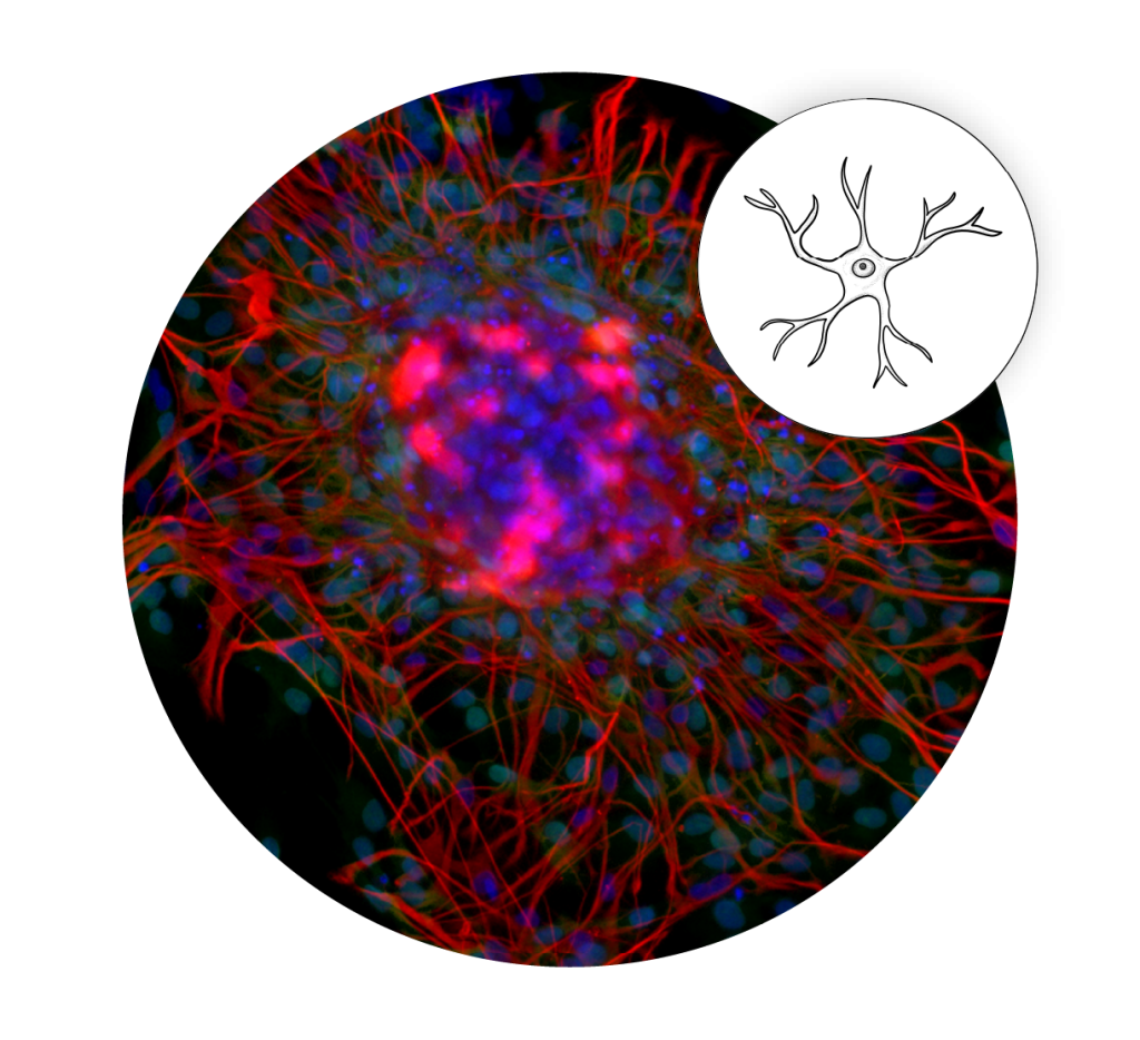
Neural Nurses: Astrocytes (Credit: Dr. Teresa Patitucci)
This image, created by Teresa Patitucci at the Medical College of Wisconsin, shows astrocytes, star-shaped cells (in red) that support the brain and form the blood-brain barrier. These particular astrocytes were grown from a patient’s skin cells using induced pluripotent stem cell technology. Since astrocytes can’t be taken directly from patients with neurological diseases like Spinal Muscular Atrophy, this approach allows researchers to study how these support cells contribute to disease development in a lab setting.

Swan Song: Human neurons (Credit: Dr. Xiaofeng Zhao)
This mirrored image of a mouse brain looks like two swans facing each other. The colors mark brain cells moving to their proper places as the brain develops. Magenta highlights migrating neurons, including those that will form the hippocampus, a brain region essential for learning and memory. This careful travel builds memory centers and other parts of the brain, which is why timing and location matter so much in development.