
Where life renews itself, one cell at a time.
Welcome to a hidden garden where cells grow, divide, and create. These images show nature’s construction zones: tubes forming, tissues regenerating, and cells branching like trees. Whether it’s the reproductive system or smooth muscle, every part of you has a plan for growth. These cells make it happen.
Try this: Which cell do you think is building something new? What might it become? Imagine this is a microscopic garden—what would you name it?
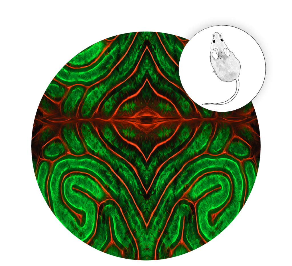
Daedalus’s Labyrinth: Reproductive tract of male mouse (Credits: Dr. Lily Oles & Dr. Kenneth Lewis)
This coiled tube in the reproductive tract of a male mouse (the epididymis) is marked by green cells lining the tubes. Here, sperm mature and are gently pushed along this network of tubules by the smooth muscle cells (in red). This maze-like structure is important for fertility and charting the twists and cell types helps understand how sperm development and movement are regulated.
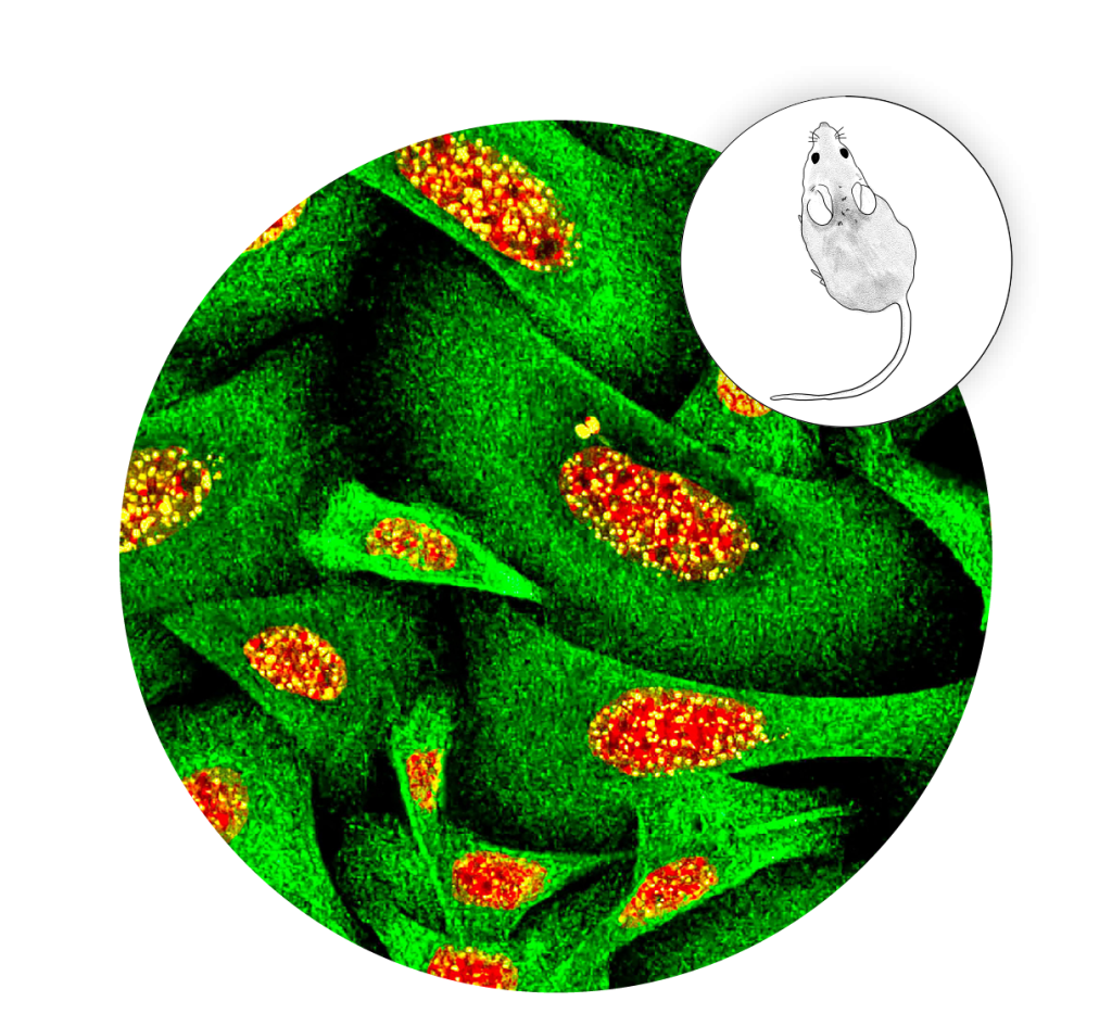
Fire in the Forrest: Mouse skin cells showing DNA damage (Credit: Dr. Raji Nair)
This image shows mouse skin cells (keratinocytes, in green) responding to DNA damage caused by radiation. Orange nuclei glow like embers while tiny yellow sparks mark a protein (gamma-H2AX) that gathers at broken DNA. This is the cell’s alarm, and the first step in repair after harmful exposure like UV light. Fast repair protects cells from changes that can cause skin cancer.
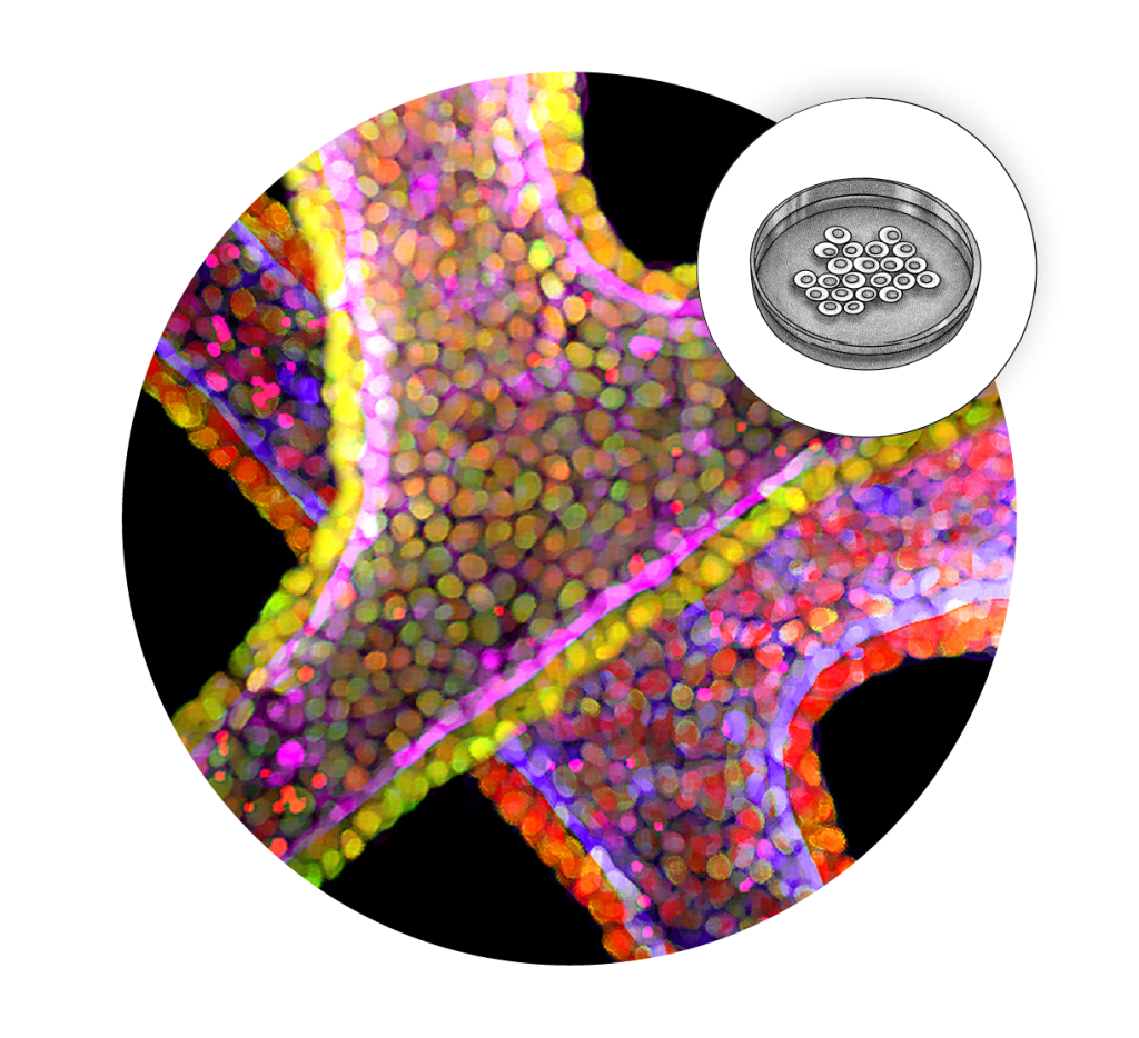
Tubing: Branched tubes formed by stem cells (Credit: Yue Shao, graduate student)
This image shows stem cells forming engineered tubes that, with the right signals, can self-organize into early shapes of organs like lungs, kidneys, and intestines, all of which rely on tubal architecture. These models teach us how organs form and how to fix them.
Smooth Operator: Engineered smooth muscle cell (Credit: Yue Shao, graduate student)
Smooth muscle powers digestion, blood flow, and more, yet many aspects of how it develops are unknown. This image shows a smooth muscle cell grown in the lab from stem cells. Creating these cells offers a new way to study smooth muscle and improve our understanding of and treatments for muscle-related diseases.
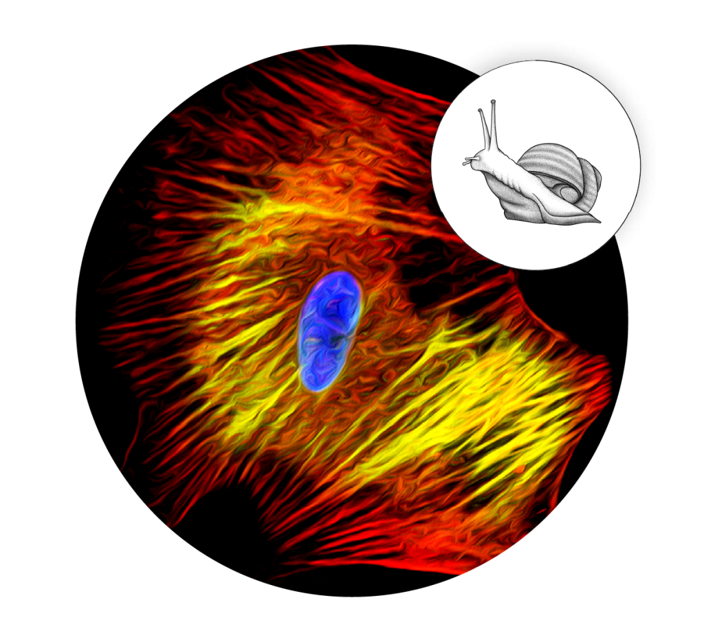
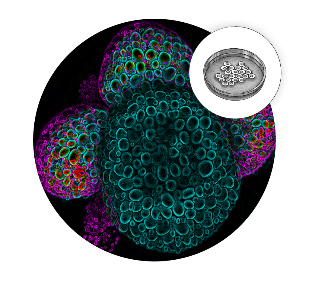
Obsoleta Beautiful: Cells of a mud snail embryo (Credit: Dr. Marcus Kilwein)
In the middle of orange and yellow fibers, eight early cells of a mud snail embryo (Tritia obsoleta) glow with alternating cyan and magenta. Each cell contains a different balance of protein (blue) and fat (magenta) which will guide what tissues these cells will become. Snail embryos provide a valuable model for understanding these processes without the ethical limitations of human embryo research.