
Two researchers from the University of Notre Dame are being honored for their outstanding publications and imaging work produced using the Integrated Imaging Facility (NDIIF). Each year, the NDIIF presents imaging awards to acknowledge and celebrate the researchers who utilize its equipment across the facility’s four Imaging Cores: Optical Microscopy, Electron Microscopy, In Vivo Imaging, and Histology.
“We are immensely grateful to the users of the NDIIF, and our annual imaging awards give us the opportunity to recognize the groundbreaking discoveries made at the facility and the dedicated researchers we collaborate with every day,” said Bradley Smith, Emil T. Hofman Professor of Science and Director of the NDIIF. “This year’s awardees exemplify the remarkable diversity and beauty of the research conducted here.” Additionally, this year was the first year recognizing one of Notre Dame’s distinguished undergraduates for his significant contributions to advancing science.
Kevin Armknecht, an undergraduate student in Prakash Nallathamby’s lab, will receive an award for best publication imaging for his research published in Nanoscale Advances in the article titled “Antimicrobial peptide-conjugated phage-mimicking nanoparticles exhibit potent bactericidal action against Streptococcus pyogenes in murine wound infection models.” Kevin is a senior majoring in Science Pre-Professional Studies with minors in Compassionate Care in Medicine and Poverty Studies.
Jarek Metro, a PhD candidate in Dr. Paul Bohn’s group, will also receive an award for best publication imaging for his research published in Analytical Chemistry titled “Nanopore-Enabled Dark-Field Digital Sensing of Nanoparticles.”
All awarded images were captured using NDIIF equipment and were taken by Notre Dame faculty, staff, or students during the 2023 calendar year. These accomplishments were celebrated at the 9th Annual Midwest Microscopy and Microanalysis Workshop on May 7th.
To learn more about the awards and the Imaging Workshop, please visit the NDIIF website.
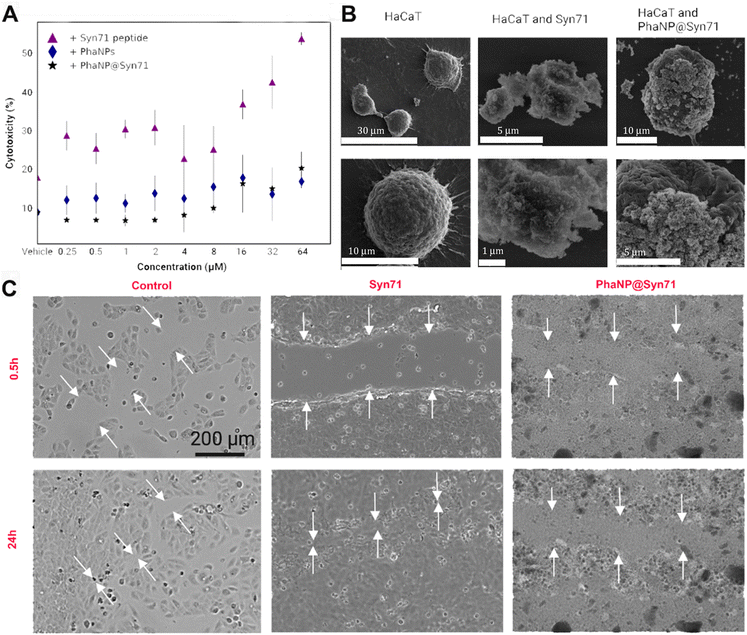
Figure Legend: (A) Cytocompatibility of PhaNP@Syn71 on the HaCaT human keratinocyte cell line. PhaNP@Syn71 were cytocompatible at all concentrations tested similar to vehicle only control. Syn71 peptide was cytocompatible to 8 μM only. The average of four replicates for each sample was computed together with standard deviations. (B) SEM images of HaCaT cells incubated with PhaNP@Syn71. This indicated that the HaCaT cells integrity was not hampered by Syn71 or PhaNP@Syn71 at 8 μM concentration w.r.t Syn71. (C) In vitro scratch closure assay on HaCaT cells showed no cell migration in the presence of PhaNP@Syn71, while Syn71 peptide only presence, or no treatment group control allowed cell migration and scratch closure. This is because the PhaNP@Syn71 had a net negative charge, which will allow them to coat the positively charged tissue culture treated gap, as a consequence of which the surface charge of the gap becomes negative and repels cells migration. The free peptides are positively charged and do not hinder cell migration. 200 μm scale bar is applicable to all optical images
Recent Comments