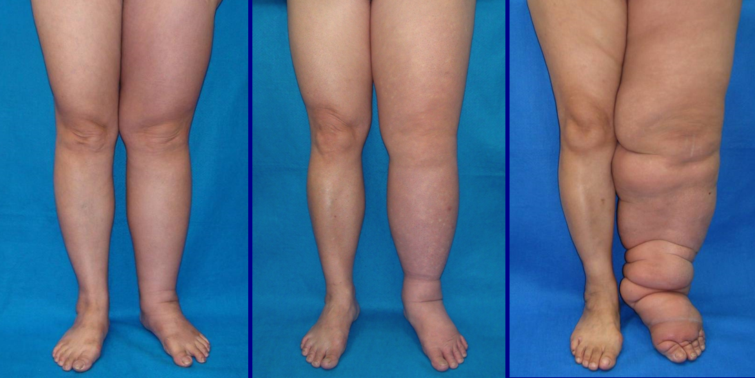
The lymphatic system is a system of vessels and lymph nodes that runs parallel to our vascular system. It takes up extra fluid from around our cells, filters it, and returns the liquid to our circulatory system. When part of the lymphatic system is damaged by surgery, radiation treatments, or injury, a progressive disease called lymphedema can occur.
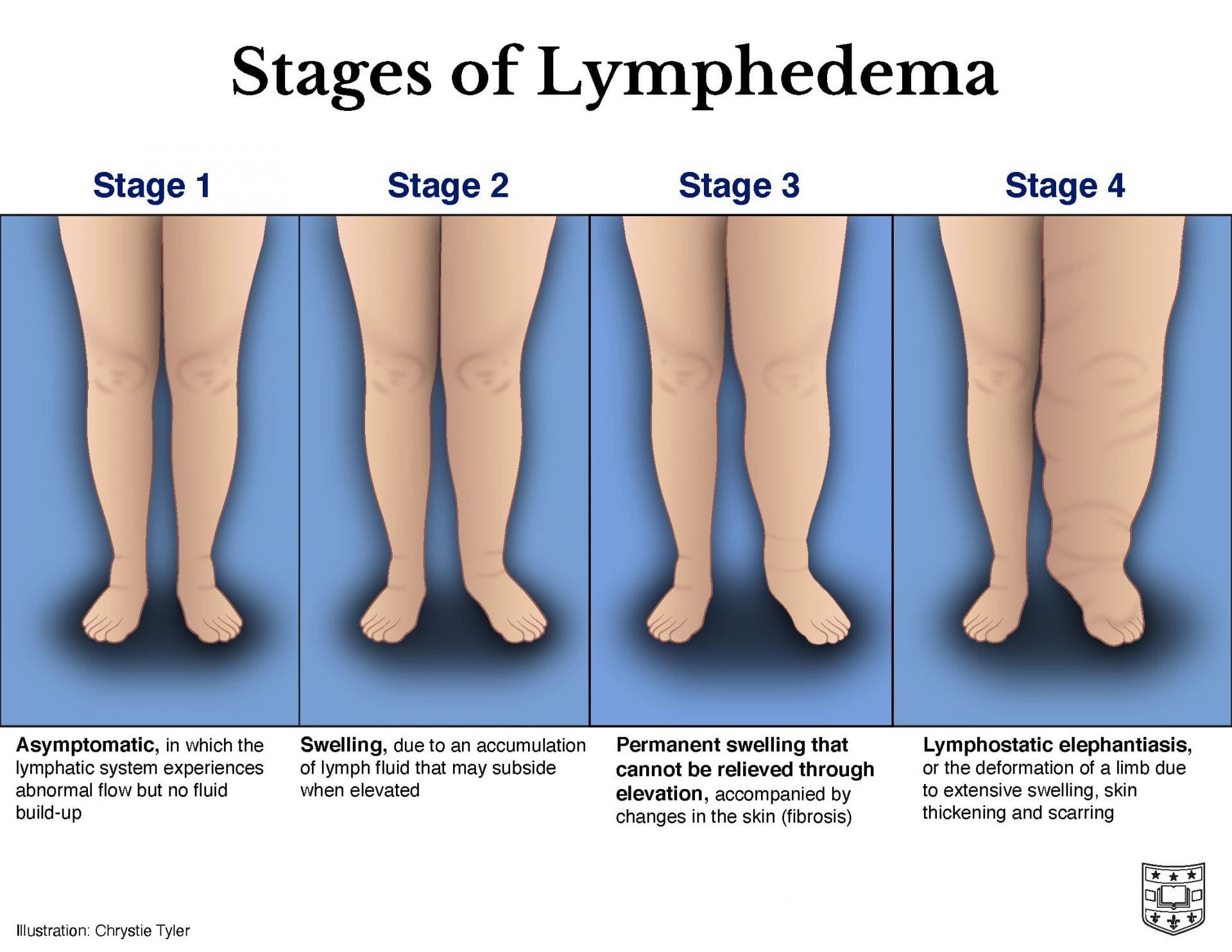
It is estimated that 20-30% of all patients who undergo lymphadenectomies during treatment for breast cancer will go on to develop lymphedema. The disease starts with swelling (edema), but it results in chronic inflammation which leads to the irreversible addition of fibrotic tissue and fat. While lymphedema can be managed in the early stages, there is currently no cure. Therefore, earlier diagnosis and consistent tracking of the disease are needed to help patients manage their condition. The following techniques can be used to non-invasively and quantitatively measure the impact of lymphedema on limb motion and tissue stiffness.
Limb motion
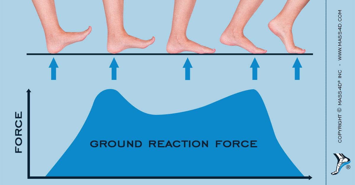
Legs: Ground reaction force measurements look at our gait as we walk. Typically, a graph will show two peaks, representing the heel and toe striking the ground, respectively. Lymphedema in the lower limb impacts how patients walk, even in the non-affected leg. Lymphedema can lead to poorer gait stability and a slower walking speed, depending on the severity of the disease.
Arms: 3D motion monitor can track the movement of our limbs in real time, giving us information about our range of motion. Researchers found that patients with severe lymphedema had limited shoulder motion compared to those with moderate lymphedema or without lymphedema.

While these techniques may not be useful to diagnose stage 1 lymphedema, it may help researchers design mobility assists and accurately track changes in movement for patients with later stages of the disease.
Tissue stiffness
Shear wave elastography measures tissue stiffness by sending a pulse through a tissue and measuring the velocity of the resulting shear wave. Just like how sound waves move faster through solid materials than liquid or air, the wave made by the pulse will move faster the stiffer a tissue is. Researchers showed that this technology could be used to non-invasively determine the levels of lymphatic obstruction in patients with lymphedema.

Extensometers pull on a material with a certain force to measure how much it stretches. By looking at the stretch of tissues over a range of forces, the stiffness of the tissue can be calculated. Here, scientists used this device to see how lymphedema changes skin anisotropy over time. Anisotropy means that a tissue has different properties in different directions. While healthy patients had no difference between the anisotropy of both arms, patients with lymphedema saw a large decrease (i.e. their skin now had a more uniform stretch instead of a one-directional stretch). Critically, this change was seen even in recently-diagnosed individuals who experienced slight swelling but no other symptoms.

While both of these techniques are useful for early diagnosis, they both rely on comparison to a healthy limb. This would make diagnosis difficult in the case of bilateral lymphedema.
Overall, mechanical testing can be used on live patients to quantify the progression of their lymphedema. While no one technique is perfect, together they open up new avenues for early diagnosis and the tracking of disease progression.
Further reading:
To explore the lymphatic system in 3D, click here!
For more information about the lymphedema, click here.
For an in-depth look on the characterization and progression of lymphedema, check out this review article. (TW: contains medical imagery. Viewer discretion advised.)