One of the leading indicators of good health is adequate lung capacity. Lung capacity, as defined by Bajaj and Delgado is the volume of air in the lungs upon the maximum effort of inspiration. For an average healthy adult, that is about 5.5 liters of air. But how do we measure our lung capacity? A spirometer is the answer. Even though the device has undergone multiple revisions since it was first invented in the 1840s, it has not deviated away from its original purpose of measuring lung capacity.
Luckily, the function of a spirometer is very intuitive to understand. One type of spirometer, called the pneumotachograph spirometer, measures the amount of air a person exhales and inhales in a second. Here is a quick run through of how that happens. The pneumotachograph spirometer typically consists of a tube, a flowmeter and a sensor. The tube is responsible for converting the information gathered by the sensor to an electric signal. The information carried by the signal is then displayed using a spirogram, a graph with flow rate (volume per second) plotted against inhaled air volume (meters cubed). Based on the characteristics of the graph, the health personnel conducting the test can then analyze the lung capacity of the subject.

Basic set-up of a spirometer test Source:Wikipedia
What are some lung conditions that a spirometer can help us diagnose? It can help us diagnose Asthma, determine if our airways have become narrowed or if our lungs are congested by mucus (pulmonary fibrosis). Another condition on the list of diagnosis is cystic fibrosis, a rare chronic condition that alters the function of body parts such as the lung and liver by producing mucus. Our vital capacities can be compromised for different reasons eventually causing the aforementioned defects in our health. Some of the reasons are partly hereditary(such as cystic fibrosis) but most of these are caused by external factors such as smoking and exposure to polluted air. Other factors cited in early medical studies include race, gender and age.
The difference in lung capacity between white people and colored people has been a widely accepted phenomenon. For a long time the broader medical community believed that lung capacity difference was innate. As a result, “race corrections” are applied on the spirometer results in an attempt to get a more accurate value. The correction factor shrinks the benchmark for standard lung capacity of black people by 10% and Asian people by 4% to 6%.
This obviously calls into question who the system designated as the benchmark of health and normalcy – the white population. The “race correction” doesn’t acknowledge the intersections of socio-economic status, exposure to cleaner air, or sex. These are all factors that can largely influence well-being including but not limited to lung capacity.
Why does this matter? It matters because race correction could result in the deprivation of the necessary medical attention that needs to be given to colored communities. It also overlooks the intersectionality of their experiences that exist in the spheres of social class, environmental factors, and lived experiences. Thus, we need to question how race correction was installed in the first place. Was it a pure speculation? Was it devised as a result of segregative policies? Or did it have an empirical basis? That is why it is important to put the spirometer in a historical context and reevaluate the implicit biases with which it was designed.
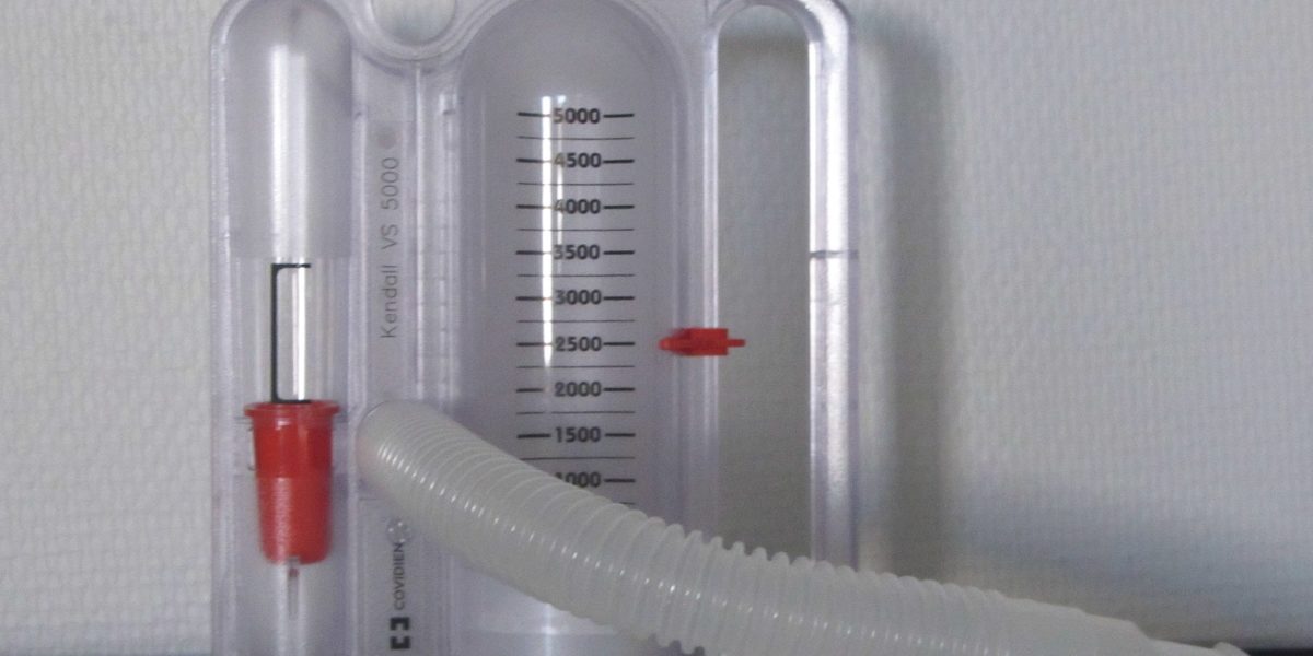
Featured image “File:Incentive spirometer2.JPG” by Stefan Bellini is marked with CC0 1.0.